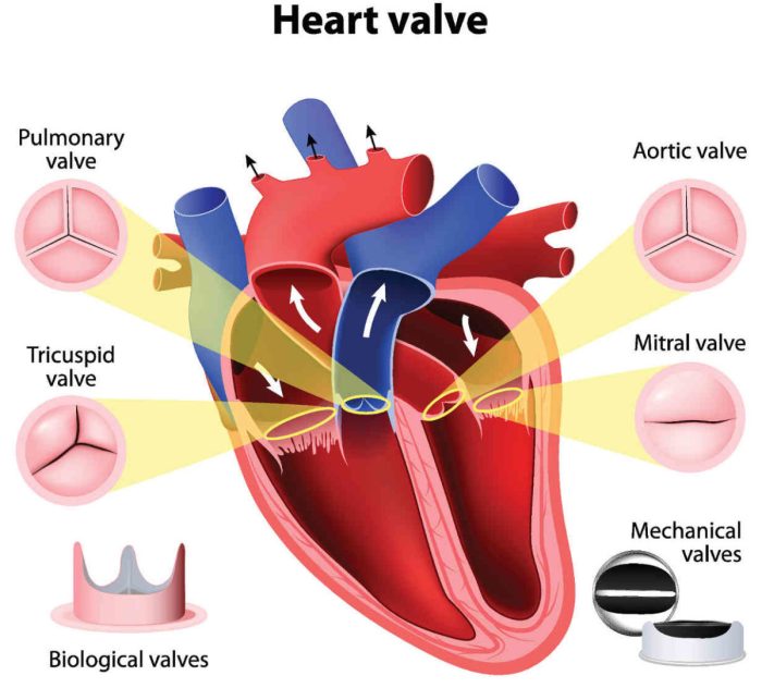
Match the following term with its correct description mitral valve – Match the following term with its correct description: mitral valve. The mitral valve, also known as the bicuspid valve, is a heart valve located between the left atrium and the left ventricle. It plays a crucial role in the cardiac cycle, preventing blood from flowing back into the left atrium during ventricular systole.
Understanding the anatomy, function, and potential disorders associated with the mitral valve is essential for comprehending cardiovascular health.
This comprehensive guide delves into the intricacies of the mitral valve, exploring its structure, function, and the various conditions that can affect its performance. We will examine the causes, symptoms, and treatment options for mitral valve disorders, providing a thorough understanding of this vital component of the human heart.
Mitral Valve Anatomy

The mitral valve, also known as the bicuspid valve, is a heart valve located between the left atrium and left ventricle. It consists of two leaflets, the anterior and posterior leaflets, which are attached to the mitral annulus by chordae tendineae.
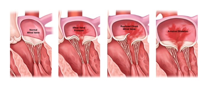
The chordae tendineae are thin, fibrous cords that prevent the leaflets from prolapsing into the atrium during ventricular systole.
The mitral valve is responsible for preventing the backflow of blood from the left ventricle into the left atrium during ventricular systole. It opens during ventricular diastole to allow blood to flow from the left atrium into the left ventricle.
Mitral Valve Function

The mitral valve plays a crucial role in the cardiac cycle. During ventricular diastole, the mitral valve opens to allow blood to flow from the left atrium into the left ventricle. As ventricular systole begins, the mitral valve closes to prevent the backflow of blood into the left atrium.
The opening and closing of the mitral valve is controlled by a complex interplay of pressure gradients, myocardial contraction, and the tension of the chordae tendineae. When the pressure in the left ventricle exceeds the pressure in the left atrium, the mitral valve closes.
When the pressure in the left atrium exceeds the pressure in the left ventricle, the mitral valve opens.
Mitral Valve Disorders
Mitral valve disorders can be classified into two main types: stenosis and regurgitation.
Mitral Valve Stenosis, Match the following term with its correct description mitral valve
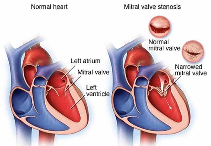
Mitral valve stenosis occurs when the mitral valve opening becomes narrowed, obstructing the flow of blood from the left atrium into the left ventricle. This can lead to symptoms such as shortness of breath, fatigue, and chest pain.
Mitral Valve Regurgitation
Mitral valve regurgitation occurs when the mitral valve does not close properly, allowing blood to leak back into the left atrium during ventricular systole. This can lead to symptoms such as shortness of breath, fatigue, and palpitations.
Mitral Valve Treatment: Match The Following Term With Its Correct Description Mitral Valve

The treatment of mitral valve disorders depends on the severity of the condition and the underlying cause. Treatment options may include:
- Medical therapy: Medications can be used to relieve symptoms and slow the progression of the disease.
- Surgery: Surgery may be necessary to repair or replace the mitral valve.
- Transcatheter interventions: Transcatheter interventions are less invasive procedures that can be used to repair or replace the mitral valve without open heart surgery.
Question Bank
What is the function of the mitral valve?
The mitral valve prevents blood from flowing back into the left atrium during ventricular systole, ensuring the proper flow of blood through the heart.
What are the common types of mitral valve disorders?
Mitral valve disorders include stenosis (narrowing of the valve) and regurgitation (leaking of the valve), both of which can disrupt the normal blood flow through the heart.
How are mitral valve disorders diagnosed?
Mitral valve disorders are typically diagnosed using echocardiography, a non-invasive imaging technique that allows doctors to visualize the structure and function of the heart valves.